
Understanding the Trochlear nerve’s unique attributes and functions is essential for medical students, particularly those specializing in neurology or ophthalmology.
As we venture further into the complexities of neuroanatomy, it becomes increasingly apparent that each cranial nerve has its own distinct characteristics and functional nuances.
The Uniqueness of the Trochlear Nerve
The trochlear nerve is distinct in several ways:
- It is the only cranial nerve that exits the brainstem dorsally.
- The smallest cranial nerve in terms of the number of axons.
- It has the longest intradural course.
Function of the Trochlear Nerve
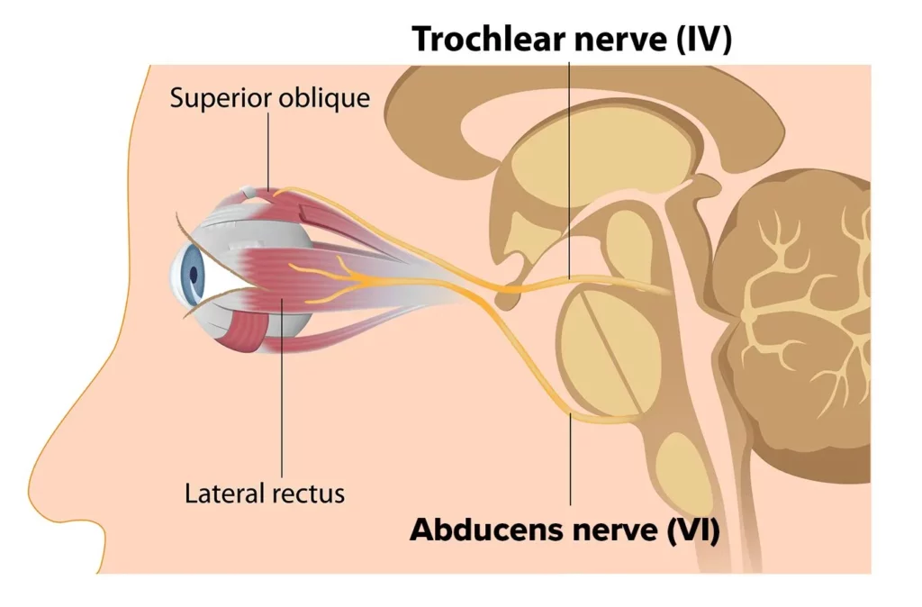
The primary function of the Trochlear nerve is to innervate the superior oblique muscle of the eye. This muscle plays a role in controlling the downward and outward movement of the eyeball, enabling intorsion, depression, and abduction of the eye.
Intracranial Route and Anatomical Relations
The trochlear nerve’s intracranial journey is quite complex and long.
Origin and Initial Course
The nerve originates from the trochlear nucleus, which is located in the dorsal aspect of the midbrain, at the level of the inferior colliculus. Unique among cranial nerves, it exits the brainstem dorsally. After emerging, the nerve fibers decussate (cross over) within the brainstem before emerging on the contralateral side. This crossing is one of the reasons for its long intradural course.
Venturing Around the Brainstem
Once it emerges, the nerve wraps around the lower border of the cerebral peduncle. This part of its pathway is a delicate maneuver, where it courses between the posterior cerebral artery and the superior cerebellar artery. This segment is particularly vulnerable to injury due to its proximity to these vascular structures.
Entry into the Subarachnoid Space
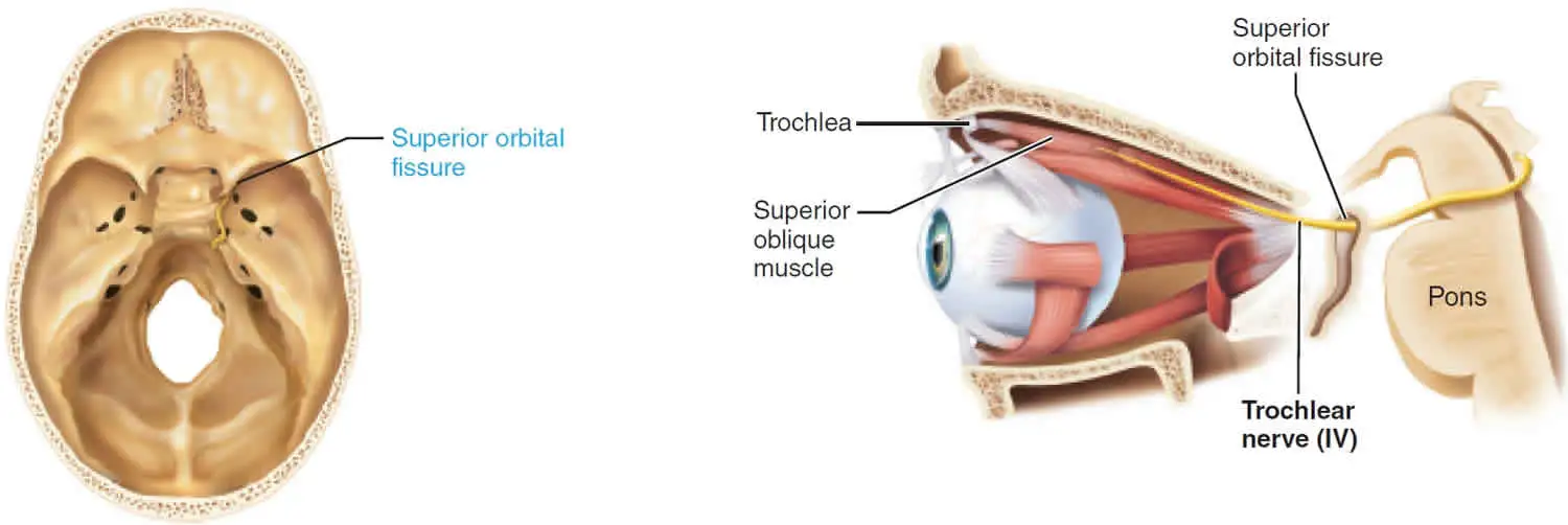
The nerve then travels forward in the subarachnoid space. It is within this space that the trochlear nerve is the longest compared to other cranial nerves. As it moves anteriorly, it lies close to the tentorium cerebelli, a dural fold separating the cerebellum from the inferior aspect of the occipital lobes.
Passage through the Cavernous Sinus
The trochlear nerve enters the cavernous sinus, a venous channel located on either side of the sella turcica. Here, it takes a superior lateral position relative to the oculomotor nerve (CN III). Within the cavernous sinus, it runs parallel to the internal carotid artery and is medial to the ophthalmic division (V1) of the trigeminal nerve (CN V).
Orbital Entry and Innervation
The nerve then proceeds to enter the orbit through the superior orbital fissure. Notably, it does so outside of the tendinous ring, which is the origin point for the rectus muscles of the eye. Finally, it innervates the superior oblique muscle, facilitating depression, intorsion, and abduction of the eye.
Clinical Relevance of the Trochlear Nerve
In the realm of clinical medicine, the trochlear nerve (CN IV) holds significant importance due to its unique anatomy and function. Its role in ocular motility makes it a critical component in the assessment of neurological and ophthalmological health.
Trochlear Nerve Palsy
This is the most common clinical presentation related to the trochlear nerve. Patients with trochlear nerve palsy experience vertical diplopia (double vision), where the images are misaligned vertically and tilted. The cause can be congenital, traumatic (as the nerve is susceptible to injury due to its long intracranial course), or secondary to microvascular diseases like diabetes.
Diagnosis often involves the Bielschowsky head tilt test, where tilting the head toward the shoulder on the side of the affected eye exacerbates the double vision.
Role in Eye Movement Disorders
Disorders like superior oblique myokymia, where there is involuntary twitching of the superior oblique muscle, can be traced back to issues with the trochlear nerve. Its unique path also makes it susceptible to compression or damage from intracranial lesions, leading to complex patterns of ocular misalignment.
Neurosurgical Implications
During neurosurgical procedures, especially those involving the brainstem or cerebellopontine angle, the Trochlear nerve’s pathway must be carefully considered to avoid inadvertent damage. Its proximity to vascular structures like the superior cerebellar artery also necessitates caution during surgical interventions.
Implications in Trauma and Neurological Disorders
Traumatic brain injuries can affect the Trochlear nerve due to its long intradural course and relative fragility. Neurological conditions like multiple sclerosis or brainstem strokes can also involve the Trochlear nerve, leading to characteristic eye movement abnormalities.
Final Words on Trochlear nerve
The trochlear nerve’s route is a masterful display of anatomical precision, illustrating the complexity of neural pathways in the human body. Its journey from the dorsal midbrain to the orbit encapsulates several critical anatomical landmarks and relationships, highlighting the importance of detailed anatomical knowledge in clinical practice.
Test your Knowledge!
Here is a brief quiz on the trochlear nerve to assess your understanding of the material covered in this article.

Study Anatomy with MedBrane
For more comprehensive anatomy quizzes, complete with detailed explanations and analytics of your simulated exams, check out the MedBrane app, available on iOS and Android.
Pingback: Understanding Hypertrophic Cardiomyopathy: A Case Study for Medical Students – MedBrane