
The brain’s dural venous sinuses, nestled within the protective layers of the dura mater, play an important role in draining blood and cerebrospinal fluid, supporting the brain’s health and proper functioning.
Understanding the Anatomy and Functions
Getting into the anatomy and function of the dural venous sinuses, we can see how these blood channels are involved in managing blood flow and stability in the brain.
Formation of dural sinuses
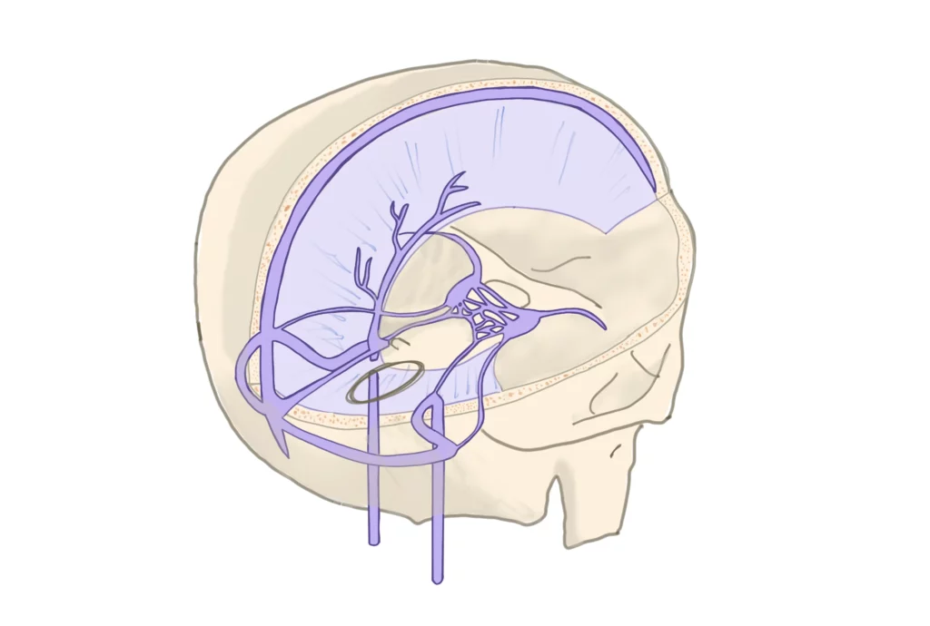
The dura mater consists of two layers: the periosteal layer, which is adherent to the inner surface of the skull, and the meningeal layer, which lies closer to the brain. The dural sinuses are formed where these two layers separate. This separation creates endothelium-lined spaces that vary in size and shape, depending on their location within the cranial cavity.
The formation of these sinuses is attributed to the folding of the dura mater, which creates partitions within the cranial cavity. These partitions are known as dural reflections or dural folds and include structures such as the falx cerebri, tentorium cerebelli, falx cerebelli, and diaphragma sellae. The dural sinuses run along these folds and at points where the dural layers come into close proximity or intersect.
Function of dural sinuses
The main function of the dural sinuses is to drain venous blood from the brain and cerebrospinal fluid from the subarachnoid space. They serve as conduits that eventually channel blood into the internal jugular veins, which carry it back to the heart.
Venous Drainage
The dural sinuses receive blood from cerebral veins, cerebellar veins, and veins of the brainstem. These veins drain blood that has circulated through the brain, carrying metabolic waste products and deoxygenated blood away from the brain tissue.
CSF Absorption
The arachnoid villi (or arachnoid granulations), which protrude into the dural sinuses, particularly the superior sagittal sinus, facilitate the absorption of CSF into the venous system. This process helps maintain the balance and pressure of CSF within the central nervous system.
Circulatory Homeostasis
By regulating the outflow of blood and CSF, the dural sinuses play a role in maintaining intracranial pressure and ensuring stable cerebral circulation. This is essential for normal brain function and the prevention of conditions such as hydrocephalus or intracranial hypertension (raised intracranial pressure).
Pathway of blood flow through the dural sinuses
Direction of Flow: Blood flow in the dural sinuses is largely governed by gravity and the pressure gradient created by cardiac activity. The flow is generally from the interior of the cranial cavity towards the base of the skull.
Venous Pressure: Unlike systemic veins, the dural sinuses do not contain valves. The pressure within the venous system and the intracranial pressure influence the rate and efficiency of blood flow through these sinuses.
Variability: There is some individual variability in the anatomy and dominance of these sinuses. For instance, one transverse sinus may be more dominant (larger) than the other.
Let’s explain the pathway of blood through the dural sinuses in 7 steps:
- Cerebral and cerebellar veins: The journey begins with smaller cerebral and cerebellar veins that drain blood from the brain’s surface and deeper structures. These veins carry deoxygenated blood and metabolic waste away from the brain tissue.
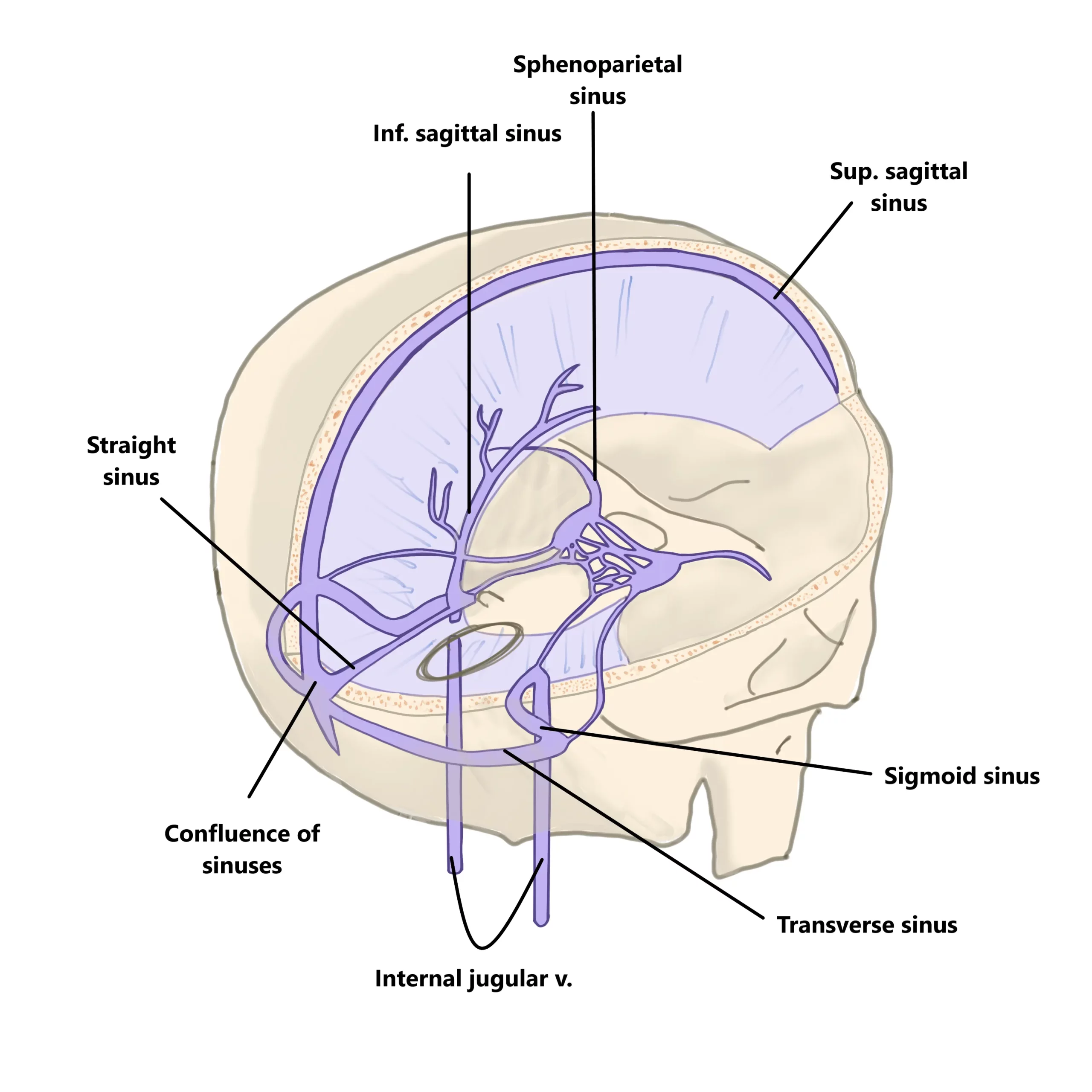
- Superior sagittal sinus: Many of these cerebral veins, particularly those from the superior aspects of the brain, empty into the superior sagittal sinus, which runs along the midline on the superior surface of the brain, within the attached edge of the falx cerebri.
- Inferior sagittal sinus and straight sinus: The inferior sagittal sinus, located in the free edge of the falx cerebri, drains blood from the medial aspects of the cerebral hemispheres and leads into the straight sinus. The straight sinus also receives blood from the great cerebral vein (of Galen), which drains the deep structures of the brain.
- Confluence of sinuses: The superior sagittal sinus, straight sinus, and occipital sinus converge at the confluence of sinuses, located near the internal occipital protuberance. This is a key juncture where blood from different parts of the brain is collected.
- Transverse sinuses: From the confluence of sinuses, blood flows laterally into the right and left transverse sinuses. These sinuses run along the attached edges of the tentorium cerebelli and curve around the posterior aspects of the cerebral hemispheres.
- Sigmoid sinuses: The transverse sinuses then continue as the sigmoid sinuses, which have an S-shaped course. They travel along the posterior cranial fossa and descend to reach the jugular foramen.
- Internal jugular veins: Finally, the sigmoid sinuses drain into the internal jugular veins, which exit the skull through the jugular foramen. The internal jugular veins carry the deoxygenated blood from the brain, along with CSF absorbed via the arachnoid villi, down the neck and towards the heart.
The cavernous sinus and clinical importance of the dural sinuses
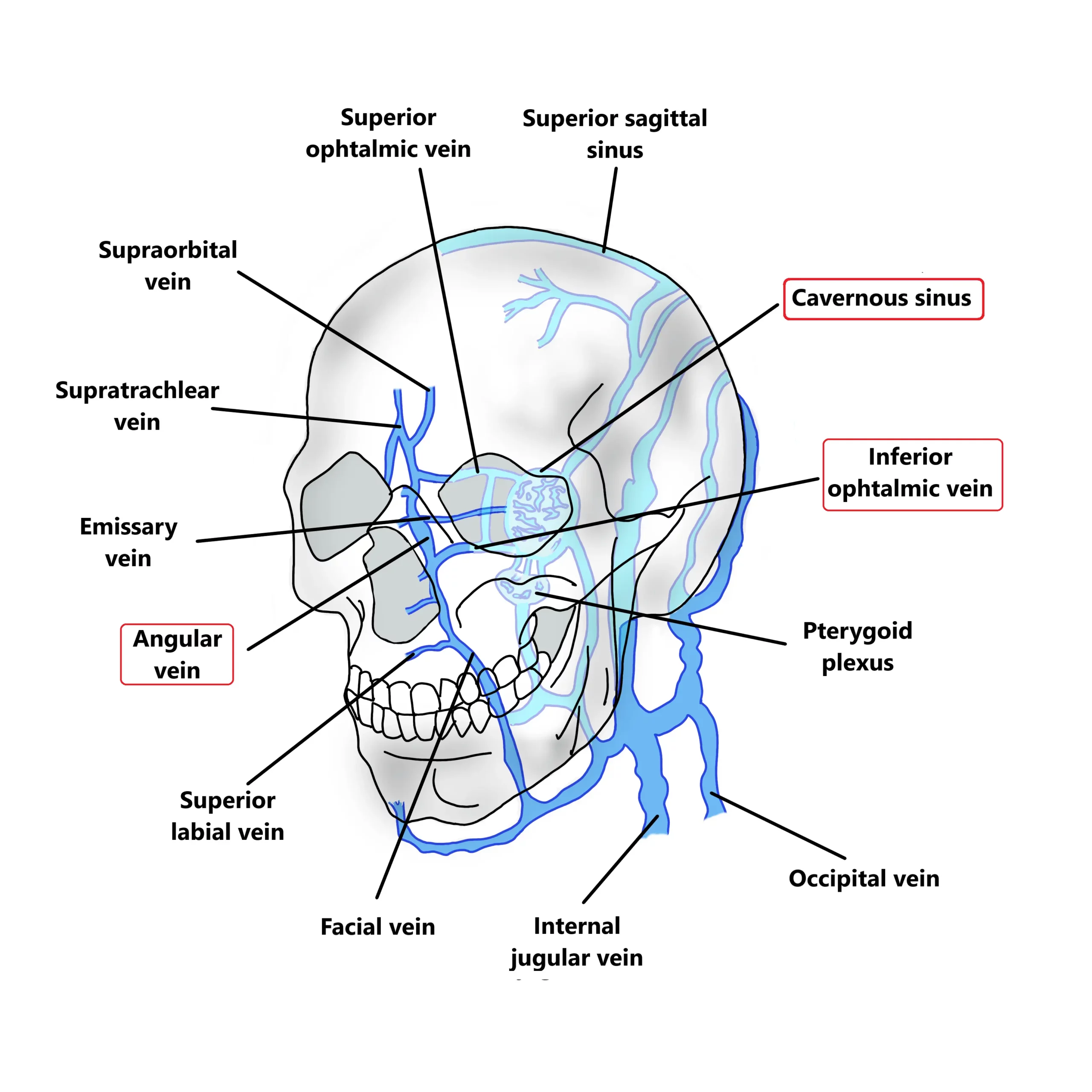
The cavernous sinus is a paired dural venous sinus situated at the base of the skull on either side of the body of the sphenoid bone. It has a unique structure, with internal carotid arteries and cranial nerves (III, IV, V1, V2, and VI) running through or in close proximity to it.
Venous Connections
Inflow
The cavernous sinus receives blood from several veins:
- Superior and inferior ophthalmic veins.
- Sphenoparietal sinus, which drains the dura mater.
- Emissary veins, which connect the sinus with the pterygoid venous plexus of the face.
- Superficial middle cerebral vein.
Outflow
Blood from the cavernous sinus drains into:
- Superior and inferior petrosal sinuses, which then drain into the transverse and sigmoid sinuses respectively.
- Pterygoid venous plexus via emissary veins.
- Inferiorly through the basilar venous plexus.
Clinical Importance of the Cavernous Sinus
The cavernous sinus is of considerable clinical significance due to its location, contents, and connections. Several aspects contribute to its clinical importance.
Cavernous Sinus Thrombosis
This is a serious condition where a blood clot forms within the cavernous sinus. It can be caused by infections spreading from the face, nose, or orbit. The angular vein, which drains the area around the root of the nose has connections to the superior ophthalmic vein which in turn drains into the cavernous sinus. The angular vein is valveless, which makes the spreading of infection from the face to the cavernous sinus easier . Symptoms of cavernous sinus thrombosis can include headache, fever, bulging eyes, and cranial nerve palsies.
Cranial Nerve Involvement
The proximity of cranial nerves III, IV, V1, V2, and VI to the cavernous sinus means that any pathology within the sinus can affect these nerves, leading to ocular motor disturbances, facial pain, or sensory loss.
Carotid-Cavernous Fistula
Abnormal communication between the internal carotid artery and the cavernous sinus can lead to increased pressure in the sinus, causing proptosis (bulging of the eye), conjunctival congestion, and potential vision loss.
General Clinical Importance of Dural Sinuses
The clinical importance of the dural sinuses extends beyond the cavernous sinus.
Thrombosis and Stroke
Thrombosis (clot formation) in any dural sinus can impair venous drainage from the brain, potentially leading to a cerebral venous sinus thrombosis (CVST). This condition can result in stroke-like symptoms, including headaches, seizures, and neurological deficits.
Intracranial Pressure
As mentioned, dural sinuses play an important role in maintaining intracranial pressure. Pathologies affecting these sinuses, like blockage or thrombosis, can lead to increased intracranial pressure, resulting in headaches, vomiting, and, in severe cases, herniation of brain tissue.
Trauma and Surgical Considerations
Injuries or surgeries near the dural sinuses carry the risk of damaging these structures, leading to bleeding or altered venous drainage, which can have significant neurological implications.

Study Anatomy with MedBrane
For more comprehensive anatomy quizzes, complete with detailed explanations and analytics of your simulated exams, check out the MedBrane app, available on iOS and Android.
Pingback: Temporal Bone: Anatomy and Clinical Perspectives | MedBrane