
The base of the skull, a complex and intricate area, has an important role in the anatomy of the head and neck area.
Serving as a bridge between the brain and the rest of the body, it not only provides support for the brain but also houses numerous anatomical structures. Let’s explore the bones constituting the base of the skull, highlight its significant features, including the sulci, grooves, and foramina, and underscore its clinical significance.
Bones and fossae of the skull base
The skull base, a complex anatomic area, comprises several bones: the ethmoid, sphenoid, frontal, occipital, and temporal. These bones form a rigid structure with multiple fossae, including the anterior, middle, and posterior cranial fossae, which house neural and vascular structures. Each fossa has distinct contents and boundaries, important for understanding cranial nerve pathways and cerebrovascular anatomy.
The bones forming the skull base
The frontal bone: This bone extends posteriorly to form the anterior cranial fossa, which supports the frontal lobes of the brain.
The Ethmoid Bone: Located at the roof of the nasal cavity, it contributes to the formation of the crista galli and cribriform plates, integral components of the middle cranial fossa.
The sphenoid bone: This butterfly-shaped bone is central to the skull’s base, contributing to all three cranial fossae and housing the sella turcica, which encloses the pituitary gland.
The temporal bone: These bilateral bones form the sides of the skull and part of the cranial floor, incorporating the middle and posterior cranial fossae.
The occipital bone: Forming the posterior skull and cranial base, it includes the foramen magnum, through which the spinal cord passes, joining the brain.
Cranial fossae
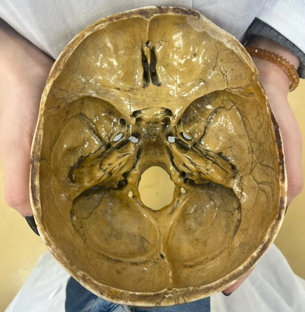
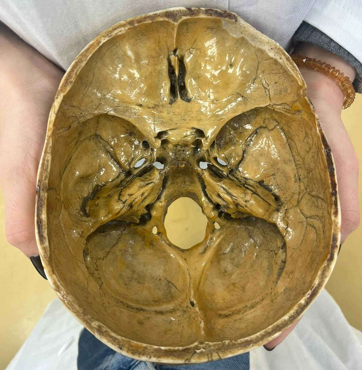
The interior of the skull base is divided into three major depressions or fossae, each conforming to the contours of the brain’s underside and accommodating different parts of the brain.
Anterior cranial fossa: The most rostral of the three, it houses the frontal lobes of the brain. The floor of this fossa is formed primarily by the frontal bone and parts of the ethmoid and sphenoid bones. The cribriform plate of the ethmoid bone, part of this fossa, contains multiple foramina for the passage of olfactory nerve fibers (CN I).
Middle cranial fossa: Deeper and situated posterior to the anterior fossa, it supports the temporal lobes and the pituitary gland. The sphenoid bone, with its sella turcica, is a central feature, providing a seat for the pituitary gland. The middle cranial fossa also contains the carotid canal for the internal carotid artery, and various foramina.
Posterior cranial fossa: The deepest fossa, accommodating the cerebellum, pons, and medulla oblongata. The occipital bone, including the foramen magnum (the largest opening in the skull), is a significant component. This fossa is crucial for the transition of the brainstem to the spinal cord and contains openings such as the internal acoustic meatus and jugular foramen for cranial nerves and the vertebral arteries.
Foramina of the skull base
The skull base has many foramina in its structure. These openings serve as passageways for various nerves and vessels. Some of the most important ones are:
The optic canal is a narrow, circular passage situated within the lesser wing of the sphenoid bone. It transmits the optic nerve (CN II) and the ophthalmic artery from the orbit to the brain.
Adjacent to the optic canal, the superior orbital fissure is a slit-like opening between the greater and lesser wings of the sphenoid bone. This fissure facilitates the passage of the oculomotor (CN III), trochlear (CN IV), abducens (CN VI) nerves, the ophthalmic branch of the trigeminal nerve (CN V₁), and the superior ophthalmic vein, all of which are integral to eye movement and sensation.
The foramen rotundum, exclusively formed by the sphenoid bone, provides a path for the maxillary nerve (CN V₂) to exit the cranial cavity. This nerve supplies sensation to the middle portion of the face.
Similarly, the foramen ovale, also part of the sphenoid bone, allows the passage of the mandibular nerve (CN V₃), the largest branch of the trigeminal nerve, which carries sensory information from the lower face and motor fibers to the muscles of mastication.
The foramen spinosum, a small opening in the sphenoid bone, is a channel for the passage of the middle meningeal artery and vein, along with the meningeal branch of the mandibular nerve, providing blood supply and innervation to the meninges.
Located at the junction between the temporal and occipital bones, the jugular foramen is larger and irregularly shaped, accommodating the internal jugular vein and cranial nerves IX (glossopharyngeal), X (vagus), and XI (accessory). These structures play a role in swallowing, taste, cardiac reflexes, and shoulder and neck movements.
The internal acoustic meatus, carved into the temporal bone, serves as a conduit for the facial (CN VII) and vestibulocochlear (CN VIII) nerves. It is crucial for hearing and balance, as well as facial expressions and taste from the anterior two-thirds of the tongue.
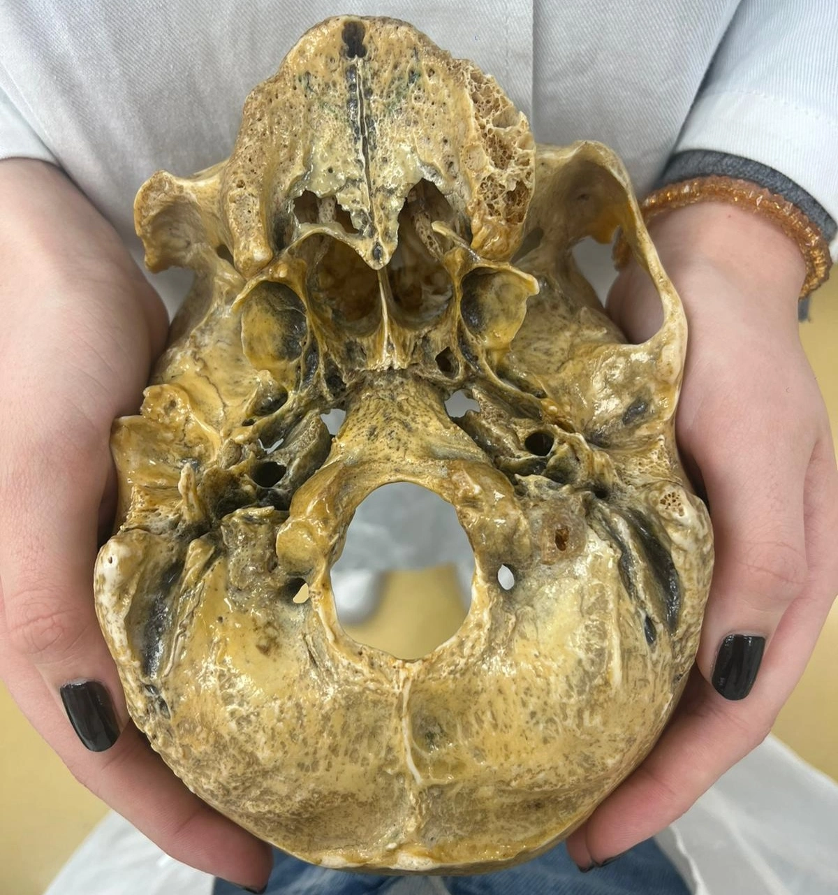
Lastly, the foramen magnum, the largest foramen located in the occipital bone, is a gateway for the spinal cord, vertebral arteries, and the accessory nerve (CN XI) to pass from the brain to the spinal column and vice versa. This foramen connects the central nervous system to the rest of the body.
Clinical importance of the skull base
Since the base of the skull serves as a connection between the skull, with its interior structures, and the rest of the body, there is a lot of important structures that can be damaged in this area.
Skull base fractures
Skull base fractures are a significant clinical concern due to the potential for severe complications. These fractures can result from blunt or penetrating head trauma and may lead to cerebrospinal fluid (CSF) leaks, meningitis, cranial nerve deficits, and vascular injuries.
The close proximity of the skull base to the brain and cranial nerves means that even minor fractures can have profound effects, necessitating prompt diagnosis and management. Clinical signs such as periorbital ecchymosis (raccoon eyes), otorrhea or rhinorrhea (CSF leak), and Battle’s sign (ecchymosis over the mastoid) can indicate skull base fractures.
Tumors of the skull base
Tumors at the skull base, including meningiomas, pituitary adenomas, chordomas, and schwannomas, present unique challenges due to their location. These tumors can compress adjacent cranial nerves or the brain itself, leading to symptoms such as visual disturbances, hormonal imbalances, hearing loss, and facial numbness or paralysis. Surgical removal of these tumors requires meticulous planning and execution to avoid damage to the critical structures housed within the skull base. Minimally invasive endoscopic techniques have revolutionized the approach to these tumors, allowing for better outcomes with reduced morbidity.
Cranial nerve disorders
The skull base’s foramina serve as passageways for the cranial nerves, making them susceptible to compression or injury from tumors, fractures, or inflammatory conditions. Conditions such as trigeminal neuralgia, hemifacial spasm, and glossopharyngeal neuralgia can arise from such compression, leading to debilitating symptoms. Microvascular decompression surgery, which involves relieving the pressure on the affected nerve, is a common treatment for these disorders and requires precise anatomical knowledge of the skull base for successful outcomes.
Advanced diagnostic and surgical techniques
The complexity of the skull base anatomy necessitates the use of advanced diagnostic techniques, including high-resolution MRI and CT scans, for accurate visualization of lesions and planning of surgical interventions. Intraoperative navigation systems and minimally invasive endoscopic approaches have significantly improved the safety and efficacy of skull base surgeries, allowing for precise targeting of pathological areas with minimal disruption to surrounding structures.
Conclusion
The skull base is crucial in the intricate anatomy of the head and neck, serving as a support for the brain and a conduit for numerous vital structures. We shed light on the constituent bones, detailed the cranial fossae, elucidated the foramina and their roles, and highlighted the clinical significance of this region. Such knowledge is indispensable for medical students, offering a foundation for understanding the complexities of skull base anatomy and its implications in health and disease.
Study with MedBrane
If you’re keen to deepen your understanding of human anatomy, give the MedBrane app a try. It’s free and offers a great way to gauge what you already know through thorough exam simulations. Plus, you can learn as you go with in-depth explanations for every question.
Also you can follow us on Instagram @medbrane for more anatomy and study tips!