The radiocarpal joint, commonly known as the wrist joint, plays an important role in the complex movements of the hand.
It is essential for medical students to understand its anatomy for accurate diagnosis and treatment of wrist injuries and pathologies.
Anatomical Structure
Bones
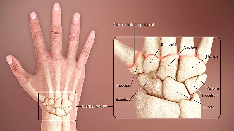
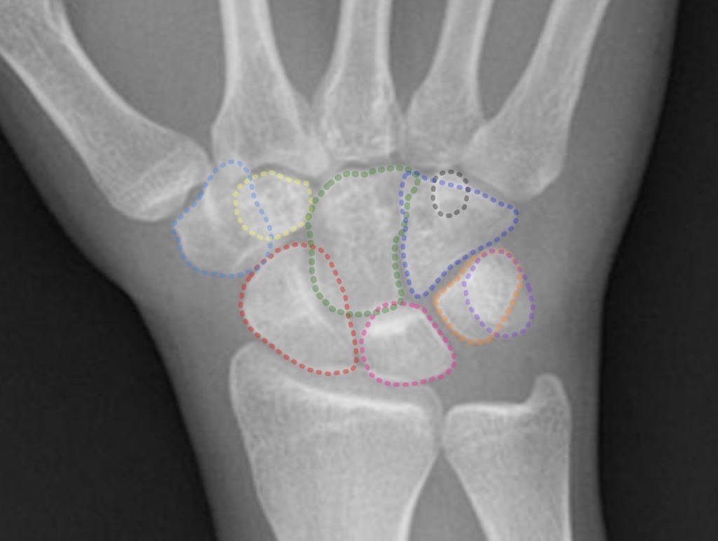
The radiocarpal joint is an ellipsoid joint, which is a type of synovial joint in which the articulating surfaces are ovoid (it is also known as a condyloid joint). The radiocarpal joint is formed by the articulation between the distal end of the radius and the proximal row of carpal bones. These carpal bones include the scaphoid, lunate, and triquetrum. The articular disc of the ulna also contributes to the joint.

Ligaments
Ligamentous structures provide stability to the radiocarpal joint. The main ligaments include:
Dorsal Radiocarpal Ligament
- Anatomy: The dorsal radiocarpal ligament is located on the back (dorsal side) of the wrist. It extends from the distal end of the radius to the carpal bones, primarily attaching to the scaphoid and lunate bones.
- Function: This ligament restricts excessive palmar flexion (bending the wrist towards the palm). It helps maintain the stability of the wrist joint during flexion and extension movements.
- Clinical Significance: Injury to this ligament can result in instability and increased palmar flexion, potentially leading to pain and decreased wrist function.
Palmar Radiocarpal Ligament
- Anatomy: Situated on the palmar side (the side of the palm) of the wrist, this ligament is broader and stronger than its dorsal counterpart. It originates from the distal radius and inserts into several carpal bones, including the scaphoid, lunate, and triquetrum.
- Function: It plays a crucial role in limiting dorsiflexion (extension of the wrist). The palmar radiocarpal ligament also contributes to the overall stability of the wrist joint.
- Clinical Significance: A damaged palmar radiocarpal ligament can result in increased wrist extension and may predispose the joint to injuries such as sprains or dislocations.
Ulnar Collateral Ligament
- Anatomy: This ligament is located on the ulnar side of the wrist, running from the styloid process of the ulna to the triquetrum and pisiform bones.
- Function: It stabilizes the ulnar side of the wrist, particularly during radial deviation (movement towards the thumb side) and helps in the distribution of load across the wrist.
- Clinical Significance: Injuries to the ulnar collateral ligament, often seen in athletes, can lead to pain, instability, and a decreased range of motion, particularly affecting movements that stress the ulnar side of the wrist.
Radial Collateral Ligament
- Anatomy: Positioned on the radial (thumb) side of the wrist, this ligament extends from the styloid process of the radius to the scaphoid bone and occasionally to the trapezium.
- Function: It provides lateral stability to the wrist, especially during ulnar deviation (movement towards the little finger side).
- Clinical Significance: Damage to the radial collateral ligament can result in lateral instability, pain, and a compromised range of motion. It’s often injured due to direct trauma or repetitive stress.
Joint Capsule
The joint capsule of the radiocarpal joint encases the joint, surrounding the articulation between the distal end of the radius and the proximal row of carpal bones (the scaphoid, lunate, and triquetrum). It is a fibrous, envelope-like structure that is lined with a synovial membrane.
The capsule attaches proximally to the margins of the distal radius and ulna, and distally to the proximal row of carpal bones. The attachment is reinforced by the intrinsic and extrinsic ligaments of the wrist.
The inner layer of the capsule is the synovial membrane, which produces synovial fluid. This fluid lubricates the joint, reducing friction and facilitating smooth movement.
Movements
The radiocarpal joint allows several types of movements, including:
- Flexion and Extension: Bending the wrist forwards and backwards.
- Radial and Ulnar Deviation: Moving the wrist towards the thumb (radial) or towards the ulna (ulnar).
- Circumduction: A circular motion that combines the above movements.
Blood Supply and Innervation
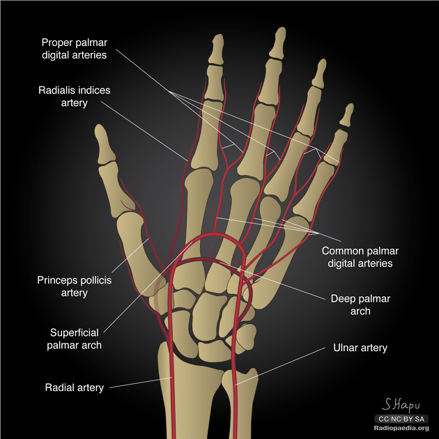
The blood supply to the radiocarpal joint is derived from a rich network of arteries, primarily the radial and ulnar arteries, which branch into smaller arterioles to extensively vascularize the area. These arteries form anastomoses (connections) around the wrist, particularly in the dorsal and palmar carpal arches, ensuring consistent blood flow to the joint and surrounding structures.
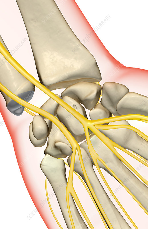
The innervation of the radiocarpal joint is complex, primarily provided by branches of the median and ulnar nerves, with some contribution from the radial nerve. These nerves supply both sensory innervation (pain and proprioceptive sensations), and motor innervation (coordinating muscles and movements of the wrist and hand). Accurate knowledge of the nerve supply is essential for diagnosing and managing wrist pathologies, as nerve injuries or compressions can lead to pain, numbness, and functional impairment in the wrist joint.

Clinical Relevance
Understanding the anatomy of the radiocarpal joint is crucial in diagnosing and treating wrist injuries such as fractures, ligament tears, and carpal instabilities. Conditions like carpal tunnel syndrome and arthritis also have implications in wrist anatomy.
Here is a brief quiz on the radiocarpal joint to assess your understanding of the material covered in this article:
Study Anatomy with MedBrane
For more comprehensive anatomy quizzes, complete with detailed explanations and analytics of your simulated exams, check out the MedBrane app, available on iOS and Android.


